


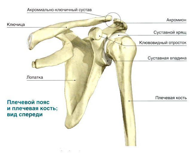
To perform the functions of support, movement and protection inour body has a system that includes bones, muscles, tendons and ligaments. All its parts grow and develop in close interaction. Their structure and properties are studied by the science of anatomy. The humerus is part of the free upper limb and along with the bones of the forearm and upper limb belt - the scapula and clavicle - provides complex mechanical movements of the human hand. In this paper, using the example of the humerus, we will study in detail the principles of the activity of the musculoskeletal system and find out how its structure is related to the functions performed.

The triangular or cylindrical shape is characteristicfor the components of the skeleton - tubular bones, in which there are elements such as the epiphyses (the edges of the bone) and its body (diaphysis). Three layers - the periosteum, the bone itself and endoost - are part of the diaphysis of the humerus. The anatomy of the free upper limb has now been studied quite well. It is known that the epiphyses contain a spongy substance, whereas the central part is represented by bone plates. They form a compact substance. This kind has long tubular bones: the shoulder, the elbow, the femur. The anatomy of the humerus, the photo of which is presented below, indicates that its shape best corresponds to the formation of mobile connections with the bones of the upper limb and forearm belt.

In the process of embryonic development, the humerustogether with the entire skeleton is formed from the middle germinal leaf - mesoderm. At the beginning of the fifth week of pregnancy, the fetus has mesenchymal areas called tabs. They grow in length and take the form of humeral tubular bones, the ossification of which continues after the birth of the child. Above, the humerus is covered with the periosteum. This is a thin shell, consisting of connective tissue and having a branched network of blood vessels and nerve endings that enter the bone itself and provide its nutrition and innervation. It is located along the entire length of the tubular bone and forms the first layer of the diaphysis. As the science of anatomy has established, the humerus, covered with the periosteum, contains fibers of the elastic protein - collagen, as well as special cells called osteoblasts and osteoclasts. They are grouped near the central channel of Havers. With age, it is filled with a yellow bone marrow.
Self-healing, repair and growth in thicknesstubular bones in the human skeleton is carried out thanks to the periosteum. Specific anatomy of the humerus in the middle part of the diaphysis. Here there is a bumpy surface, to which a superficial deltoid muscle joins. Together with the upper limb belt and the bones of the shoulder and forearm, it provides lifting and retraction of elbows and hands up, back and in front of you.
The extremities of the tubular bone of the shoulder are calledepiphyses, contain red bone marrow and consist of a spongy substance. His cells produce blood cells - platelets and erythrocytes. Epiphyses are covered with the periosteum, they have bone plates and cords called trabeculae. They are located at an angle to each other and constitute the inner skeleton in the form of a system of cavities, which are filled with a hematopoietic tissue. As the anatomy has determined, the structure of the humerus at the junction with the shoulder blade and the bones of the forearm is quite complicated. The articular surfaces of the humerus have proximal and distal ends. The head of the bone has a convex surface covered with hyaline cartilage and entering the cavity of the scapula. Special cartilaginous formation of the scapula - an articulate lip - serves as a shock absorber, which softens shocks and impacts when the shoulder moves. The capsule of the shoulder joint is attached at one end to the scapula, and the other to the head of the humerus, sinking to its neck. It stabilizes the connection between the shoulder girdle and the free upper limb.
As the human anatomy has established, the humerusis included in the composition not only of the spherical shoulder joint, but also one more - a complex ulnar joint. It should be noted that the shoulder joint is the most mobile in the human body. This is understandable, since the hand serves as the main tool of labor operations, and its mobility is associated with the adaptation to the upright and release from participation in the movement.
The elbow joint consists of three separatejoints connected by a common joint capsule. The distal humerus is connected to the ulnar bone, forming a block-shaped joint. Simultaneously, the head of the humerus of the humerus enters the fossa of the proximal end of the radius, forming a brachial mobile connection.
The normal anatomy of the humerus includesa large and a small apophyses - the tubercles, from which the crests leave. They serve as a place of attachment of the muscles of the shoulder. Here is a groove, which serves as a receptacle for the biceps tendon. At the border with the bone, diaphysis, below the apophyses, there is a surgical neck. It is most vulnerable to traumatic shoulder injuries - dislocations and fractures. In the middle of the body of the bone there is a knobby region to which the deltoid muscle is attached, and behind it is a furrow of a spiral shape into which the radial nerve is immersed. At the boundary of the epiphyses and diaphysis lies the site, the rapidly dividing cells of which cause the growth of the humerus in length.
The most common injury isfracture of the shoulder with a fall or strong mechanical shock. The reason lies in the fact that the joint does not have real ligaments and is stabilized only by the muscular corset of the upper extremity belt and the auxiliary ligament, which looks like a bundle of collagen fibrils. Suffer common lesions of the soft tissues of the shoulder joint, for example, tendonitis and capsulitis. In the first case, the tendon of the supraspinous, subacute, and small round muscles is damaged. Another disease occurs as a result of inflammatory processes in the joint capsule of the shoulder.

Pathologies are accompanied by tunnel pain inarm and shoulder, limiting the mobility of the shoulder joint with the lifting of the arms upwards, setting them behind their backs, leading them to the sides. All these symptoms dramatically reduce the efficiency and physical activity of a person.
In this article, we studied the anatomical structure of the humerus and found out its relationship with the functions performed.

